The peripheral nervous system consists of nerves that branch out from your central nervous system into the body's main mass. (Waxenbaum et al. 2019) It relays information from your brain and spinal cord to your organs, arms, legs, fingers and toes and it consists of two distinct and separate evolutionary systems; the more recent somatic nervous system, which guides your voluntary movements and an evolutionary much older system, the autonomic nervous system, which controls many of the activities we do without us having to think consciously about them. The autonomic nervous system regulates certain body processes, such as blood pressure and the rate of breathing and it helps regulate the internal organs, including the blood vessels, stomach, intestine, liver, kidneys, bladder, genitals, lungs, pupils, heart, as well as sweat, salivary, and digestive glands, in response to both internal and external messages about the changing state of the environment. It has two response divisions; the sympathetic and the parasympathetic. When the autonomic nervous system receives new information about the body and/or the external environment, it responds by stimulating body processes, usually through the sympathetic division, or inhibiting them, usually through the parasympathetic division. The communication system involves a nerve pathway whereby one cell is located in a main processing section of the nervous system, the brain stem or spinal cord and another is located in a cluster of nerve cells (called an autonomic ganglion), which are connected with the various internal organs that need controlling. So by either stimulating or inhibiting these organs in doing what they do, the system controls internal body processes such as blood pressure, heart and breathing rates, body temperature, digestion, metabolism, chemical balances, production of body fluids, urination, defecation and sexual responses. I.e. most of what is going on to sustain our daily lives. For example, when the sympathetic division acts on the heart and blood vessels it increases blood pressure, whilst messages from the parasympathetic division decreases it. The two divisions work together to ensure that the body responds appropriately to different situations. Because we don't see how this works, we have to imagine what it might be like. Just as the changing echo sounds of a submarine sonar system might be listened to in order to visualise the shape of a nearby whale, imaginative images based on stories told in order to illicit representations of the feeling tones of someone's body might be used to develop images of what unseen things might look like.
In 'The Strange Order of Things' by Antonio Domasio, the idea of neural mapping is introduced.
'At some point, long after nervous systems were able to respond to many features of the objects and movements that they sensed, both inside and outside their own organism, there began the ability to map the objects and events being sensed. This meant that rather than merely helping detect stimuli and respond suitably, nervous systems literally began drawing maps of the configurations of objects and events in space, using the activity of nerve cells in a layout of neural circuits. .........imagine the neurons wired in circuits and laid out on a flat board, where every point of the surface corresponds to a neuron. Then imagine that when a neuron in the circuitry becomes active, it lights up, something akin to making a dot on a board using a marker. The ordered, gradual addition of many such dots generates lines that can link up or intersect and create a map'. (2019, p. 76)
Because we are so good at creating these imaginative maps of what we cant see, we have externalised the skills, so that we can build all sorts of cognitive maps in our minds that relate to external perceived experiences. A cognitive map is a type of mental representation that helps us to acquire, store, recall and decode information about the relative locations and attributes of experiences found in both real and/or metaphorical or invented spatial environments. Cognitive mapping then gives us a parallel process whereby we know where we are in space and how that location is related to other locations. Mapping can also add value to these places, such as where the best places to eat are or what areas to avoid because they are dangerous. What the implications of this mean are that before we made that very basic first drawn map, perhaps one that was scratched out using a stick in soft earth, we had developed the capacity to map out those relationships inside our body/minds. Not only that, but we had also developed representations of pain location as well as feeling tone. For instance a tightness in the chest might be due to stress or a lightheadedness due to sexual excitement, but we in some way knew where that tightness was located. These feelings are with us from the moment our nervous systems begin to operate. Therefore not only is the body's map known and used as a model before we begin to map out the environment it inhabits, but as the body grows an awareness of its environment, the map it builds is one based on its initial cognitive mapping of itself, which includes emotional awareness.
There has been some evidence for the idea that cognitive maps are represented in more than one way. (Jacobs et al 2003) One is the bearing map, which represents the environment through self-movement cues. These are vector-based cues that create a rough, 2D map of the environment. Another is a sort of sketch map that works using positional cues. The second map integrates specific objects, or landmarks and their relative locations to also create a 2D map of the environment. The integration of these two separate maps with other sources of information, such as memories of touch or awareness of the body, gradually shapes a growing awareness of the environments that people inhabit. Our bodies are very simple in many ways, they are bilaterally symmetrical and we have our main sense organs located at one end, therefore body maps can have a left / right, top / bottom structure and we apply this to the external world as well. What particularly interests myself is that scientists believe that individual place cells within the hippocampus correspond to separate locations in the environment, with the sum of all cells contributing to a single map of an entire environment. The strength of the connections built up between the cells represents the distances between them in the actual environment. In effect our physical structure is amended and reshaped in response to experiences, just as a map on paper you make or draw from personal experience, is composed of a constant re-drawing and editing in order to get a closer and closer account of the information you gather.
Related, but in many ways very different, are the Body Maps that are often used by medical practitioners to visually pinpoint where on the body a medical condition is affecting a patient. This helps them accurately track the physical progression of an illness so that care can be applied accordingly. A wide selection of conditions can be recorded whereby changes to the surface area of the map over time are important, such as bruises, scalds, infections, skin conditions or swellings as well as being a way to give precision to letting others know where a broken bone or pain may be, or the site of a previous injection. These maps tend to be outlines of the front and back of a male or female human. You can get them in adult and child size, but all the parts of the figure are related in a 'normalised' position and no accounts are made of the different psychological or or internal maps made by individuals, in these cases 'one shape fits all'.
Body map. Head (area 1, 2, 23, or 24); neck (area 3 or 25), shoulders (area 4, 5, 26, 27); arms (area 6, 7, 8, 9, 28, 29, 30 or 31); hand (area 10, 11, 32, 33); ribs or chest (area 12 or 13); abdomen (area 14 or 15), back (area 34, 35, 36, 37), buttocks or hips (area 38 or 39); genitalia (area 16), legs (area 17, 18, 19, 20, 40, 41, 42 or 43); feet (area 21, 22, 44 or 45). Adapted from Margolis, Tait, & Krause (1986).
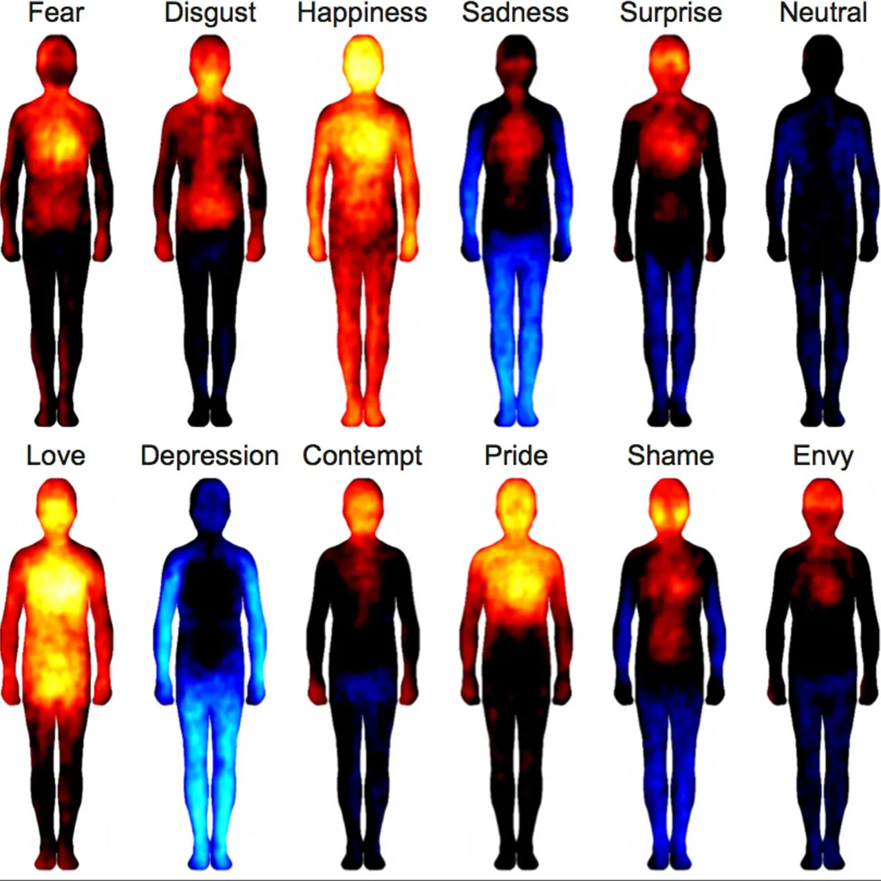
However if we look at diagrams of how we actually sense the world, we find that the body is sensed very differently to the two front and back figure drawings above.
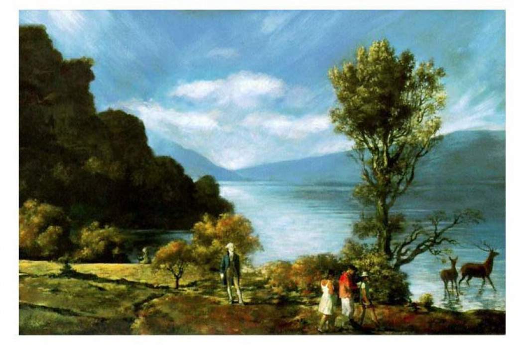
The standard human shape in the diagram above is for many people acceptable but the colour range feels far too predictable, but I suppose if you are trying to communicate across several cultural barriers, then a certain crudity of emotion / colour correspondence is to be expected. It does though seem to me to be all too neat, it looks like a carefully designed answer, rather than the confused mess of colour, shape and emotion that I have had to deal with when I have spoken to people about visualising their emotional worlds. I'm reminded of Vitaly Komar and Alexander Melamid's work whereby they statistically researched what each country's population thought would be a most loved and most hated painting, and then making visible the results. The final body of work was very carefully designed and illustrated the points they wanted to make very well, but once you 'got it', you 'got it' and that seemed a long long way away from the actual experience of encountering either a painting or a landscape.
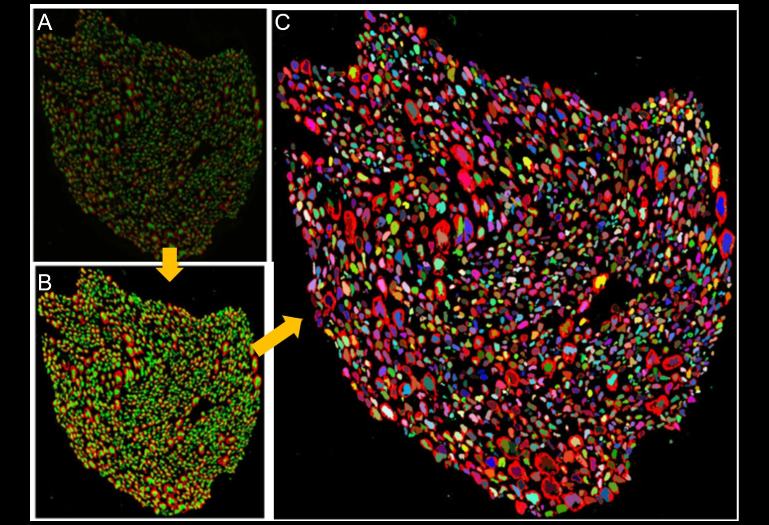
The map will show the anatomical connectivity of vagus nerve fibres from the brainstem, where the vagus nerve starts, all the way down to organs in the neck, chest and abdomen. At the moment how sensory and motor fibres are arranged inside the vagus nerve and pathways to different organs are essentially unknown. The vagus nerve, which means “wandering” in Latin, runs from the brainstem to most of the body’s major organs. The nerve is not one solid structure but is instead made up of more than 100,000 sensory and motor fibres, grouped together in several bundles, or fascicles, inside the nerve, eventually forming branches that connect to the organs like the heart, lungs, oesophagus, liver and intestines. When functioning properly, the vagus nerve contributes to the maintenance of our body’s homeostasis. However, diseases like Crohn’s disease and rheumatoid arthritis, chronic heart diseases, diabetes and even some cancers are associated with inflammation and abnormal vagus nerve function. This research has already provided images that I could imagine at some point feeding into my own thinking about how the body has developed its own maps. The process of mapping being nested, going from initial maps made in the mind to these new ones that will rely on computer enhancement and the techniques of electron microscopy. The thinking behind this project emerging from minds structured in particular ways that allow them to think of ideas in certain ways, ways that are always responses to information being processed internally and externally; this two way process reflecting the fact that the vagus nerve itself is transmitting information in both directions between the brain and body.
Barrett, L.F., 2017. How emotions are made: The secret life of the brain. London: Pan Macmillan.
Damasio, A., 2019. The strange order of things: Life, feeling, and the making of cultures. London: Vintage.
Downs, R. M. and Stea, D. 1977 Maps in Minds: Reflections on cognitive mapping London: Harper and Row
Godfrey-Smith, P. 2021 Metazoa London: William Collins
Volynets, S., Glerean, E., Hietanen, J. K., Hari, R., & Nummenmaa, L. 2020 Bodily maps of emotions are culturally universal. Emotion, 20(7), 1127–1136. https://doi.org/10.1037/emo0000624
Jacobs, F. 2022 Satirical cartography: a century of American humor in twisted maps Big Think Available at: https://bigthink.com/strange-maps/satirical-cartography-9th-avenue-steinberg/
Jacobs, Lucia F.; Schenk, Françoise (April 2003). Unpacking the cognitive map: the parallel map theory of hippocampal function. Psychological Review. 110 (2): 285–315.Libassi, M. (2022) Feinstein awarded $6.7M NIH grant to create first human vagus nerve anatomical map Northwell Health: Newsroom, October 25th, Available at: https://www.northwell.edu/news/the-latest/6-7m-nih-grant-creates-first-human-vagus-nerve-anatomical-map
See also:
Drawing analogue and digital processes How certain visualisations evolved from making images of internal body sensations


Thanks for sharing your great views on this blog. The impact of technology on the education sector is always an exciting thing to inspect. The article "2025 must have educational apps to use each teacher in 2025" includes information of great equipment for teachers. As an education app development company believes that innovation can improve a lot about how people learn. Continue to share helpful resources, as everyone really values them.
ReplyDelete